Introduction: To treat patients with advanced chronic or before end stage liver diseases (hepatitis B, C, D, autoimmune chronic hepatitis, cirrhosis, variceal bleeding, low platelet count), ideally tissues or organs transplantation are needed. However, this type of intervention is severely limited by lack of donors. In this context, the culture of hepatocytes on polymeric scaffolds to generate a three-dimensional tissue was developed during the past years [1, 2]. The polymeric matrices or scaffolds must possess appropriate: i) porosity and interconnective pore network for mass transport and cellular organization in space; ii) biocompatibility and a certain bioresorbability; iii) a suitable surface for cell attachment, proliferation, differentiation; iv) adequate mechanical properties similar to those of natural organs. For such applications, porous structures with pore size ranging between 50 and 200 μm (adequate for hepatocyte culture) are required and in some cases chemical modification of the polymer is required to promote cell adhesion. We are now proposing a new series of hepatocyte functionalized supports based on polyvinyl alcohol (PVA) cross-linked by sodium trimetaphosphate (STMP) using an original two step method.
Material and Methods: A volume of 100 mL of PVA (10 % solution) was first cross-linked with sodium trimetaphosphate (STMP) under two-speed steering, until a foam was formed. Then, 1mL of NaOH (30 %) was added and the foam was molded into 6 well plates followed by freeze-drying. Derivatized scaffolds were prepared in the same way and conditions also adding galactose, collagen, chitosan or modified starch. Scaffold strips were placed into a 12 well plates and seeded with isolated Rat (Sprague-Dawley) hepatocytes at a density of 2 x 10⁶ cells/strip.
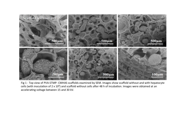
Results and discussions: The proposed method produces high porous matrices (≥ 70 %) with a porosity distribution between 50 and 1000 μm (scanning electron microscope [SEM] micrographs - Fig 1). This reorganization of the cells inside the pores looked strangely to tissue like structures. Smaller and medium-sized pores are able to retain hepatocytes, facilitating their ability to interact and adhere to the support. The PVA scaffolds show hepatocyte adhesion superior to 80 % and viability of more than 50 % (superior to the values reported for the two-dimension [2D] cell cultures). The addition of a derivatization agent does not improve attachment except in the case of chitosan-derivatized scaffold when the hepatocyte retention was affected by the presence of chitosan. High efficiency of cellular adhesion in the 3D PVA microenvironment was also shown by our group (2) in a long-term dynamic culture of hepatocytes.
Conclusion: The advantage of using a 3D microenvironment for cell culture lies in the beneficial influence that this structure has on hepatocyte behavior. In 3D system the support increases the “physical” interactions between the membrane proteins of hepatocytes and the matrix whereas for 2D cultures, only one surface of the cells is involved which may play a critical role in cell aggregation. This hepatocyte seeded scaffolds can be potentially used for liver tissue regeneration and for current evaluation of various drugs and toxic agents.
Acknowledgement: NSERC-CRSNG Innov and Discovery programs for supporting this research
References:
[1] Wang X, Yan Y, Lin F, Xiong Z, Wu R, Zhang R, Lu Q. Preparation and characterization of collagen/chitosan/hparin matrix for an implantable bioartificial liver. J Biomater Sci Polymer Edn 2005; 16: 1063-1080.
[2] Saavedra YGL, Mateescu MA, Averill-Bates DA, Denizeau F. Polyvinylalcohol three-dimensional matrices for improved long-term dynamic culture of hepatocytes. J Biomed Mater Res A 2003; 66: 562-570.