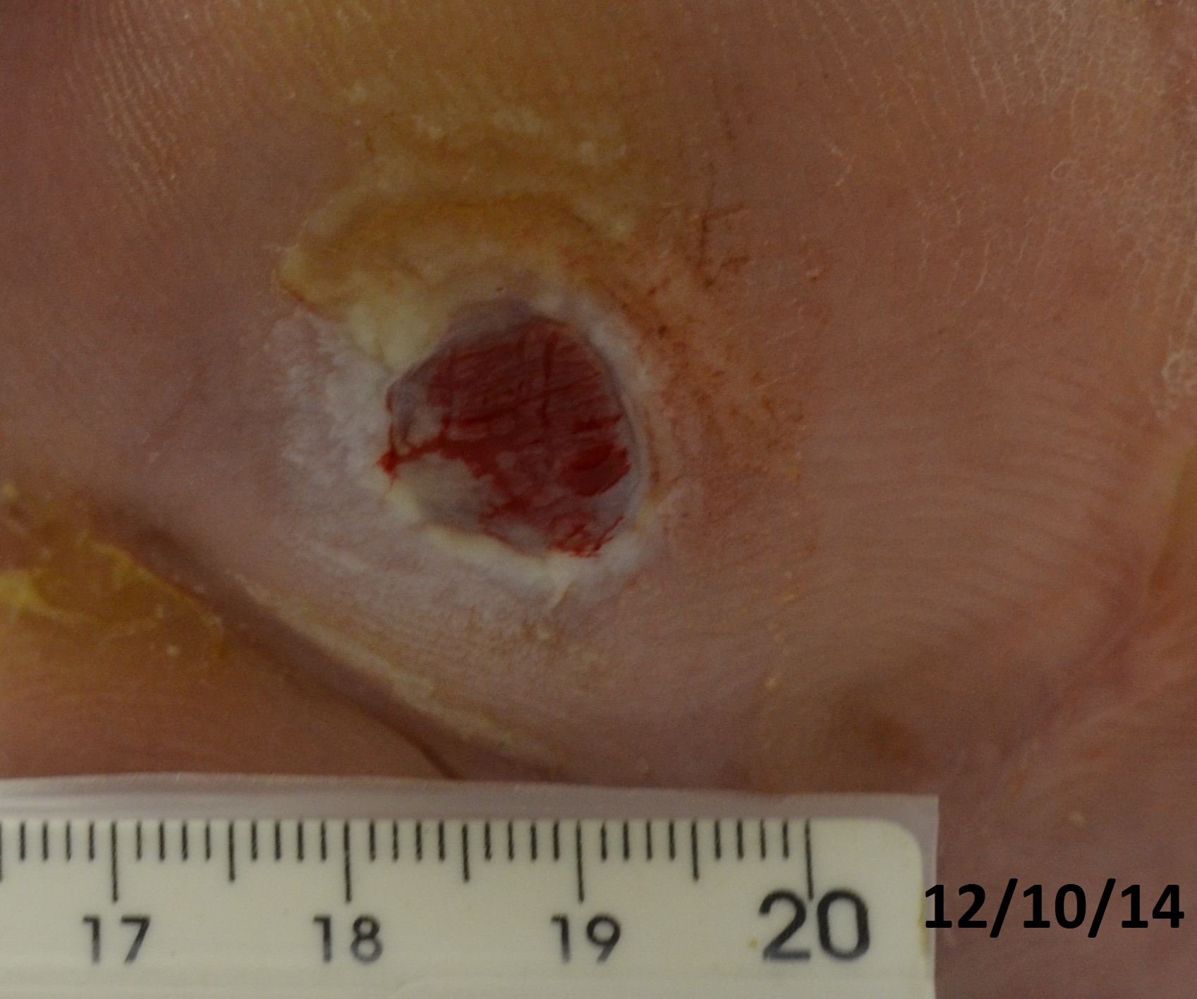
Introduction: Diabetes mellitus (DM) is an important worldwide public health problem, affecting 5–10% of the population, with an estimated prevalence in 2035 of almost 600 million people (Sweitzer, et al., 2006; Bakker, et al., 2015). The quality of life of these individuals is impaired due to the significant resulting complications, such as foot ulcers, with an incidence of 2-4% in developed countries (Bakker, et al., 2015). Foot ulcers are among the main complications of DM, being characterized by difficult healing when treated with conventional methods. Frequently, amputation of lower limbs is necessary, with great social and economic repercussion. In this work, a 61 years old man, with an amputation of the 4th and 5th right foot toes and an open plantar ulcer not healed for one year of about 1 cm in width, and a 50 years old man, with a 7 month interdigital ulcer caused by local trauma which resulted in local osteomyelitis had their lesions treated through an approach involving autologous cells and different types of biopolymers.
Methodology: Both patients had a history of 25 years of DM and underwent previous conventional skin lesion treatments, without healing. After approval of proper institutional ethics committee, they were submitted to a cell culture therapy that consisted of culturing autologous fibroblasts from skin biopsies followed by direct cell application to the surface of the ulcers with fibrin glue and subsequent coating with a chitosan-xanthan film, without previous debridement. The autologous fibroblast cultures were obtained from the dermis and cultured in the presence of specific culture medium (Rehder et al., 2004; 2013). The chitosan-xanthan dressing was produced by complexing the polysaccharides at controlled conditions of polymer addition rate, mixing, complexation and drying temperature and properly characterized prior to use (Bellini et al., 2012; 2015). Follow-ups occurred weekly for 1 month, and then twice a month. The size and aspect of the lesions and the general condition of the patients were analyzed.
Results and Discussion: Less than 12 hours after the beginning of the therapy, both patients reported pain cessation. A few days later, intense exudation attributed to fibrin, granulation tissue and the presence of serous crusts in the ulcers were detected, with subsequent centripetal closure of the wound in one patient and centrifugal closure in the other. The ulcers were fully healed by the end of the 1st month in one patient and in 5 weeks in the other one. After 9 months of follow up, the ulcers remained healed. No adverse reactions derived from the treatment.


Conclusion: The autologous fibroblast cell therapy coupled with chitosan-xanthan films demonstrated to be an effective and fast approach to treat diabetic foot ulcers, with great functional and esthetical results. Reduced healing time of diabetic ulcers was achieved, even for lesions associated with osteomyelitis.
Fundação de Amparo à Pesquisa do Estado de São Paulo (FAPESP, Brazil); Conselho Nacional de Desenvolvimento Científico e Tecnológico (CNPq, Brazil); Coordenação de Aperfeiçoamento de Pessoal de Nível Superior (CAPES, Brazil)
References:
[1] Bakker K, Apelqvist J, Lipsky BA, et al. 2015; The 2015 IWGDF Guidance Documents on Prevention and Management of Foot Problems in Diabetes: Development of an Evidence-Based Global Consensus Diabetes Metab Res Rev. doi: 10.1002/dmrr.2694
[2] Bellini MZ, Caliari-Oliveira C, Mizukami A et al. 2015; Combining xanthan and chitosan membranes to multipotent mesenchymal stromal cells as bioactive dressings for dermo-epidermal wounds. Journal of Biomaterials Applications 29(8): 1155–1166
[3] Bellini MZ, Pires ALR, Vasconcelos MO et al. 2012; Comparison of the properties of compacted and porous lamellar chitosan-xanthan membranes as dressings and scaffolds for the treatment of skin lesions. Journal of Applied Polymer Science 125: E421–E431
[4] Rehder J, Bosnardo CAF, Leite MBP et al. 2013 ; A comparative study of cell therapy and fibrin glue applied to chronic venous ulcers. Procedia Engineering 59 : 85-91
[5] Rehder J, Souto LR, Issa CM et al. 2004 ; Model of human epidermis reconstructed in vitro with keratinocytes and melanocytes on dead de-epidermized human dermis. São Paulo Med J 122(1):22-5
[6] Sweitzer SM, Fann SA, Borg TK, et al. 2006; What is the future of diabetic wound care? The Diabetes Educator 32(2):197–210