Behaviour of hMSC cultured on piezoelectric quasi-3D substrates: growth and osteogenic differentiation
Rita
Almeida1, 2,
Maria Noel
Tamaño-Machiavello1,
Leonor
Senent3*,
Lourdes
Cordón3*,
Daniela
M.
Correia2*,
Clarisse
Ribeiro2*,
Senentxu
Lanceros-Méndez2*,
Roser
Sabater I Serra1*,
Amparo
Sempere3* and
Jose Luis
Gomez Ribelles1, 4*
-
1
Universitat Politècnica de València, Center for Biomaterials and Tissue Engineering, Spain
-
2
Universidade do Minho, Centro/Departamento de Fisica, Portugal
-
3
Hospital Universitari i Politècnic La Fe, Servicio de Hematología, Spain
-
4
Biomedical Research Networking Center in Bioengineering, Biomaterials and Nanomedicine (CIBER-BBN), Spain
Introduction: Poly(vinylidenefluoride) (PVDF) is a biocompatible piezoelectric polymer that can adopt different crystalline phases: the α-phase is formed by crystallization from the melt in a broad range of temperatures and shows no piezoelectricity while the electroactive β-phase can be obtained by deformation of an α-phase PVDF film, by casting from specific solvents and/or introducing specific fillers. In previous works pre-osteoblastic cell culture on PVDF substrates has shown the effect of electroactive phase polarization on cell attachment and proliferation and a significant effect of electro-mechanical stimulation on the evolution of cells in culture[1],[2]. In this work we analyze the combined effect of piezoelectricity a surface topography on the growth and differentiation of human mesenchymal stem cells.
Methods: PVDF microspheres were produced by electrospray[3] resulting in a polymer with a high concentration of β-phase. The microspheres were projected on α-phase PVDF films during the electrospray process yielding surfaces with varying surface density of microspheres that were used as substrates for cell culture (Figure 1 shows SEM images of hMSCs cultured on supports with high (a) or low (b) density of PVDF microspheres). Flat α and β-phase films were used as reference. Human mesenchymal stem cells hMSC were seeded on the substrates and characteristic cell surface markers of hMSC’s (CD105, CD90 and CD73) were analyzed by flow cytometry (FC) before and after the different culture conditions both in expansion culture medium (until confluence) or in osteogenic medium (after confluence). Proliferation was assessed by MTS after 4 days. Cell morphology was assessed by Scanning Electron Microscopy (SEM).
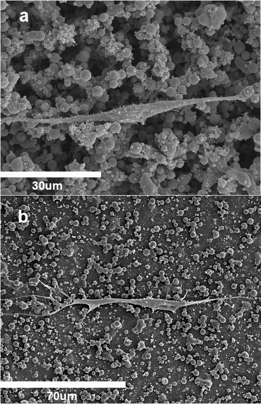
Results: Cell seeded on the microspheres substrates find an environment somway intermediate between monolayer and 3D, as shown in Figure 1 hMSCs tend to adopt enlarged morphologies. No significant differences were found in the proliferation of hMSCs in the supports with different topography. Interestingly, the expression of characteristic markers of multi-potentiality diminished during the first four days in expansion medium in a way that depends on the surface density of β-phase microparticles. The expression of osteogenic markers was also assessed in the different supports after culture in osteogenic medium.
Conclusions: Electroactive PVDF supports hMSCs culture favouring osteogenic differentiation. Cells adhere to the rough substrate and even in expansion medium this topography induces loss of multi-potentiality in hMSCs before reaching confluence.
JLGR, DMC and CR gratefully acknowledge the financial support from the Spanish Ministry of Economy and Competitiveness through the MAT2013-46467-C4-1-R project, including FEDER funds.; CIBER-BBN is an initiative funded by the VI National R&D&I Plan 2008-2011, Iniciativa Ingenio 2010, Consolider Program, CIBER Actions and financed by the Instituto de Salud Carlos III with the assistance from the European Regional Development Fund.; CR and DMC would like to acknowledge the FCT for the SFRH/BPD/90870/2012 and SFRH/BD/82411/2011 grants, respectively.
References:
[1] C. Ribeiro, J.A. Panadero, V. Sencadas, S. Lanceros-Méndez, M.N. Tamaño, D. Moratal, M. Salmerón-Sánchez, J.L. Gómez Fibronectin adsorption and cell response on electroactive poly(vinylidene fluoride) films. Biomedical Materials 7, 035004-14 (2012)
[2] C. Ribeiro, S. Moreira, V. Correia, V. Sencadas, J.G. Rocha, F. M. Gama, J.L. Gómez Ribelles and S. Lanceros-Méndez Enhanced proliferation of pre-osteoblastic cells by dynamic piezoelectric stimulation RSC Advances 2(30) 11504-11509 (2012)
[3] D. M. Correia, R. Gonçalves, C. Ribeiro, V. Sencadas, G. Botelho, J. L. Gomez Ribelles, S. Lanceros-Méndez Electrosprayed poly(vinylidene fluoride) microspheres for tissue engineering applications. RSC Advances 4, 33013-33021 (2014)
Keywords:
Cell Differentiation,
stem cell,
biomaterial,
surface topolography
Conference:
10th World Biomaterials Congress, Montréal, Canada, 17 May - 22 May, 2016.
Presentation Type:
New Frontier Oral
Topic:
Biomimetic materials
Citation:
Almeida
R,
Tamaño-Machiavello
M,
Senent
L,
Cordón
L,
Correia
DM,
Ribeiro
C,
Lanceros-Méndez
S,
Sabater I Serra
R,
Sempere
A and
Gomez Ribelles
J
(2016). Behaviour of hMSC cultured on piezoelectric quasi-3D substrates: growth and osteogenic differentiation.
Front. Bioeng. Biotechnol.
Conference Abstract:
10th World Biomaterials Congress.
doi: 10.3389/conf.FBIOE.2016.01.02160
Copyright:
The abstracts in this collection have not been subject to any Frontiers peer review or checks, and are not endorsed by Frontiers.
They are made available through the Frontiers publishing platform as a service to conference organizers and presenters.
The copyright in the individual abstracts is owned by the author of each abstract or his/her employer unless otherwise stated.
Each abstract, as well as the collection of abstracts, are published under a Creative Commons CC-BY 4.0 (attribution) licence (https://creativecommons.org/licenses/by/4.0/) and may thus be reproduced, translated, adapted and be the subject of derivative works provided the authors and Frontiers are attributed.
For Frontiers’ terms and conditions please see https://www.frontiersin.org/legal/terms-and-conditions.
Received:
27 Mar 2016;
Published Online:
30 Mar 2016.
*
Correspondence:
Dr. Leonor Senent, Hospital Universitari i Politècnic La Fe, Servicio de Hematología, Valencia, Spain, senent_leo@gva.es
Dr. Lourdes Cordón, Hospital Universitari i Politècnic La Fe, Servicio de Hematología, Valencia, Spain, lou.cordon@gmail.com
Dr. Daniela M Correia, Universidade do Minho, Centro/Departamento de Fisica, Braga, Portugal, danielamscorreia@gmail.com
Dr. Clarisse Ribeiro, Universidade do Minho, Centro/Departamento de Fisica, Braga, Portugal, clarisse.engbiom@gmail.com
Dr. Senentxu Lanceros-Méndez, Universidade do Minho, Centro/Departamento de Fisica, Braga, Portugal, lanceros@fisica.uminho.pt
Dr. Roser Sabater I Serra, Universitat Politècnica de València, Center for Biomaterials and Tissue Engineering, Valencia, Spain, rsabater@die.upv.es
Dr. Amparo Sempere, Hospital Universitari i Politècnic La Fe, Servicio de Hematología, Valencia, Spain, sempere_amp@gva.es
Dr. Jose Luis Gomez Ribelles, Universitat Politècnica de València, Center for Biomaterials and Tissue Engineering, Valencia, Spain, Email1