Introduction: Glioblastoma multiforme (GBM), the most malignant brain tumor, are highly invasive and frequently exploit microvessels as guides for migration. Hence, interfering with perivascular invasion of GBM cells is promising to add a new therapeutic direction to reduce glioblastoma dispersion, but the lack of proper 3-D vascular niche model restricts investigating cell-cell interaction between glioblastoma and vasculature using patient-derived cancer cells. In this study, we adapted an interdisciplinary approach combining patient-derived GBM cells and vascular bioengineering using 3-D bioprinting technology[1]-[3] to create a model of glioblastoma-vascular niche.
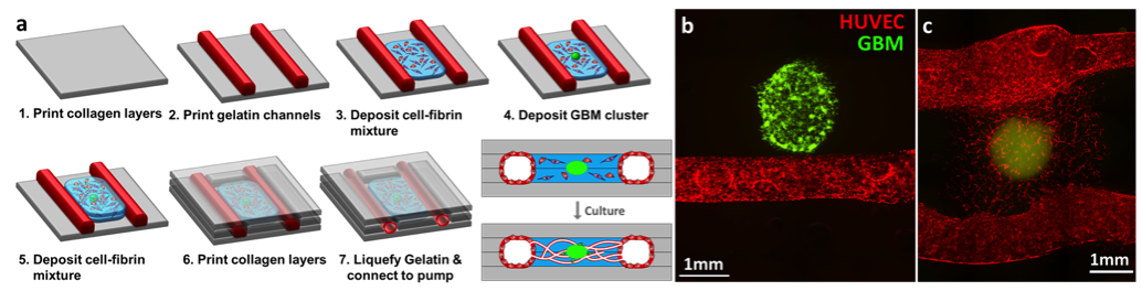
Materials and Methods: We first investigated GBM invasion pattern in different matrix compositions in order to define optimal scaffold condition for the GBM-vascular niche model. GBM cell clusters were embedded within the collagen I matrices with different concentration of laminin (0, 10, 100 µg/mL) or hyaluronic acid (HA) (0-10 wt%). To fabricate 3-D GBM-vascular model, fluidic vascular channels were created within 3D collagen I matrix. For GBM-capillary niche, fibrinogen (10 mg/mL), thrombin, human umbilical vein endothelial cells (HUVECs), and fibroblasts (FBs) were deposited in between the two channels during the fabrication process. GBM cell clusters were located nearby the vascular channels or within the capillary bed (Fig. 1a).
Results and Discussion: In the invasion test, GBM cells were embedded within collagen scaffolds and cultured for 7 days. The average invasion distance for collagen matrices with 0, 10, 100 µg/mL laminin were 443±134 µm, 599±189µm, and 706±78µm, respectively. The invasion distance was also increased with 10wt % of HA (635±119µm). We developed two types of GBM-vascular niche model. The first model which consists of one large fluidic vessel and adjacent GBM cluster was developed to study angiogenesis and vessel co-option by GBM cells (Fig. 1b). The embedded GBM cells proliferated, invaded towards the outer collagen scaffold, and reached the edge of vascular channel after 7 days of culture. The second model contains capillary bed connected to large vessels for the purpose of modeling interaction of GBM cells with microvasculature (Fig. 1a). More active capillary formation was observed around the GBM cluster (Fig. 1c).
Conclusions: This study shifts the research focus to an important area of tumor vasculature microenvironment and utilizes cutting-edge 3D bio-printing technology to establish innovative 3-D glioblastoma-vascular systems with dynamic flow to model GBM-vasculature interactions. By real-time observation of cell-cell interactions in 3D glioma/vascular systems, our studies will lay the foundation for novel therapeutic development to curtail glioma invasion and improve GBM therapy. The 3-D vascular niche platform can also be easily adapted to other biological system and will serve as a unique tool to model stem cell interaction with vascular niches.
This work was supported by NIH R01HL118245, NSF CBET 1263455, NSF Career 1350240.
References:
[1] Lee VK, Lanzi AM, Haygan N, Yoo SS, Vincent PA and Dai G. Generation of Multi-Scale Vascular Network System within 3D Hydrogel using 3D Bio-Printing Technology. Cell Mol Bioeng. 2014;7:460-472.
[2] Lee VK, Kim DY, Ngo H, Lee Y, Seo L, Yoo SS, Vincent PA and Dai G. Creating perfused functional vascular channels using 3D bio-printing technology. Biomaterials. 2014;35:8092-102.
[3] Zhao L, Lee VK, Yoo SS, Dai G and Intes X. The integration of 3-D cell printing and mesoscopic fluorescence molecular tomography of vascular constructs within thick hydrogel scaffolds. Biomaterials. 2012;33:5325-32.