Introduction: The concept of multifunctional devices that combine drug eluting components together with functional prosthetic implants, represents an emerging clinical technology that promises to provide functional enhancement to implant devices in various clinical applications[1],[2]. The properly designed drug delivery system can allow for a precise drug release control, which refers to the amount of released dose, the drug release profile and the speed of release[3]. In general, the choice of the proper drug loading technic can be decisive for obtaining the suitable and desired drug release from biomaterial[4]. The aim of this work was to compare different methods of the drug loading into chitosan/ polyurethane matrix in terms of optimization of the developed systems.
Materials and Methods: In the first stage chitosan microspheres were prepared using emulsification/cross-linking method[5]. Chitosan solution in acidic acid containing gentamicin (3 or 6 wt. % vs. chitosan mass) was emulsified in the dispersion medium consisting of liquid paraffin stabilized by the sorbitan monooleate. Triphosphate pentasodium (TPP) and glutaraldehyde (25 or 50 wt.% solution in H2O) were used as a cross-linking agents. Next the microspheres loaded with gentamicin were added to the synthesis of polyurethane matrix (PU). PU networks based on three-branched hydroxyl terminated caprolactone (CL) and glycolide-co-lactide (PLGA) oligomers were synthesized using 1,6-hexamethylene diisocyanate (HMDI) and dibutyltin dilaurate as the catalyst. Additionally, polyurethanes consisting of chitosan macromolecule segment were synthesized and drugs were incorporated directly into the polymer matrix during synthesis. In vitro release study was performed after immersion of the composites in PBS according to the method used by Zang[6].
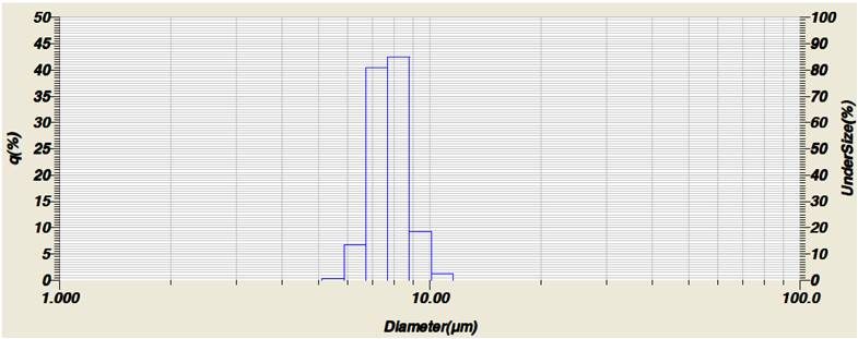
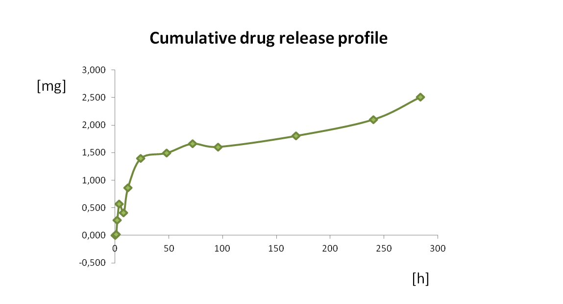
Results and Discussion: Two types of biodegradable polyurethane composites containing chitosan microspheres or chitosan macromolecule segments were obtained. Chitosan microspheres with mean diameter of 8 μm (Fig.1) were distributed within polymer matrix. Drug loading efficiency in case of microspheres system increased significantly form 3% for microspheres crosslinked with 50 wt.% glutaraldehyde to 60% for microspheres crosslinked with 25 wt%. glutaraldehyde. Drug release profile from the gentamicin containing-microspheres composites proved to be stable and sustained for more than a month (Fig. 2.). In case of the polyurethane composites containing the microspheres with the drug loaded directly to the polymer matrix, the higher burst release was observed due to the facilitated diffusion of the drug.


Conclusion: The results of drug release study showed that the drug release kinetics from the chitosan/polyurethane composites system can be modified by drug loading method and the cross-linking degree of chitosan microspheres. The study confirms that these systems could be successfully used un regenerative medicine as drug carriers.
This work was financed by National Science Centre on the basis of a decision number DEC- 2012/07/D/ST8/02588.
References:
[1] MEH El-Sayed, A. S. Hoffman, P.S. Stayton, Smart polymeric carriers for enhanced intracellular delivery of therapeutic macromolecules, Expert Opinion on Biological Therapy 5(1) (2005) 23-32.
[2] M.R.Aguilar, C. Elvira, A. Gallardo, B. Vázquez, and J.S. Román. Smart polymers and Their applications as biomaterials.
[3] A.K. Bajpai ∗, S. K. Shukla, S. Bhanu, S. Kankane. Responsive polymers in controlled drug delivery. Progress in Polymer Science 2008;11(33): 1088–1118
[4] C. Wischke, A. T. Neffe, S. Steuer, A. Lendlein. Comparing techniques for drug loading of shape-memory polymer networks--effect on their functionalities. Eur J Pharm Sci. 2010 Sep 11;41(1):136-47.
[5] E. Szymańska, K. Winnicka, M. Muśko. Otrzymywanie mikrosfer chitozanowych metodą emulsyjno-sieciującą z zastosowaniem trifosforanu (TPP) I aldehydu glutarowego (GLT).
[6] Zhang X, Wyss UP, Pichora D, Goosen MFA. Biodegradable controlled antibiotic release devices for osteomyelitis: optimization of release properties. J Pharm Pharmacol 1994;46:718–24.