Introduction: The interface between bone and ligament or tendon (enthesis) is characterized by a multiphasic interface containing spatial gradients in mechanical properties, structure and cell type. These gradients exist over a distance of less than one millimeter and serve to mediate load transfer between highly dissimilar tissues by minimizing stress concentrations. As mechanical mediators in the musculoskeletal system, the insertion sites of ligaments or tendons with bone have high incidences of acute and long-term injury respectively[1]. Current surgical repair for many of these tissues is largely unsatisfactory as it does not regenerate the complex structure of the natural interface[2]. Given the variety and prevalence of failures, repair or regeneration of interface tissues remains a significant clinical challenge. In order to satisfactorily regenerate the proper bone-tendon interface, graded tissue engineering scaffolds will be required that properly mimic natural interface mechanical properties. To achieve this it is essential to first understand the chemical and mechanical changes across the natural enthesis. This study focuses on the mechanical transition measured using image correlated tensile testing alongside finite element modelling.
Materials and Methods: Shoulders from skeletally mature porcine were dissected to leave the humeral head, supraspinatus tendon and 2 cm of supraspinatus muscle. The supraspinatus insertion site complex was then frozen in liquid nitrogen and sectioned into 1 mm thick slices. Samples were thawed for testing and split along the length of the tendon. These were placed in a miniature loading machine (Ernest F Fullam, Latham, NY, USA), speckled & placed under an upright optical microscope (BX-51 M, Olympus, Markham, Canada). Strains were calculated using Digital Image Correlation (DIC) with the commercial software VIC-2D (Correlated Solutions, SC, USA). Finite element modelling (FE) of 2D tendon-bone junctions was conducted in Abaqus (Dassault Systems, Vilacoublay, France).
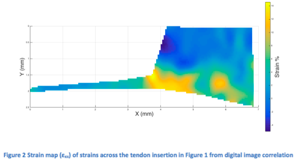
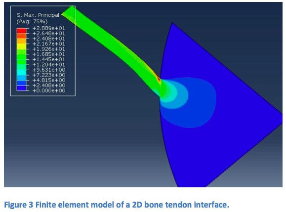
Results and Discussion: Tensile testing of the tendon-bone junction (Figure 1) was used with DIC to generate uniaxial strain maps (Figure 2), revealing a stress concentration at the acute corner of the enthesis. This is in line with theoretical predictions of the material junction of these dissimilar tissues[3] and with the observations of the FE model (Figure 3). By varying the location, width and profile of the transition in modulus of the FE simulated interface the natural modulus transition in these tissues can be inferred. It is observed that the natural enthesis does not seem to employ a lower modulus transition region as has been suggested[4],[5].

Conclusions: The combination of DIC analysis of tensile tests of tendon-bone junctions along with FE models of a 2D structure allows substantial insight into the mechanics of the natural tendon bone interface. On a material level it is clear stress concentrations are an inevitability of this system and hence any strategies to reduce them must be on a macroscopic, geometrical level.
This work has been developed as a result of a mobility stay funded by the Erasmus Mundus Programme of the European Commission under the Transatlantic Partnership for Excellence in Engineering – TEE Project.; This research was supported by an EPSRC DTA award to O.E.Armitage
References:
[1] United States Bone & Joint Initative, The Burden of Musculoskeletal Diseases in the United States, Second Edition. Rosemont, IL: American Academy of Orthopedic Surgeons, 2011.
[2] S. A. Rodeo, S. P. Arnoczky, P. A. Torzilli, C. Hidaka, and R. F. Warren, “Tendon-healing in a bone tunnel. A biomechanical and histological study in the dog.,” J. Bone Jt. Surg., vol. 75, no. 12, pp. 1795–1803, 1993.
[3] G. M. Genin and Y. Liu, “Models for the Mechanics of Joining Dissimilar Materials,” in Structural Interfaces and Attachments in Biology, G. M. Genin, S. Thomopoulos, and V. Birman, Eds. 2013, pp. 43–68.
[4] Y. X. Liu, S. Thomopoulos, V. Birman, J. S. Li, and G. M. Genin, “Bi-material attachment through a compliant interfacial system at the tendon-to-bone insertion site,” Mech. Mater., vol. 44, pp. 83–92, 2012.
[5] G. M. Genin, A. Kent, V. Birman, B. Wopenka, J. D. Pasteris, P. J. Marquez, and S. Thomopoulos, “Functional Grading of Mineral and Collagen in the Attachment of Tendon to Bone,” Biophys. J., vol. 97, pp. 976–985, 2009.